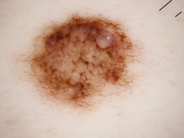
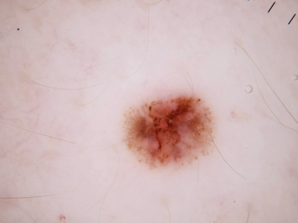
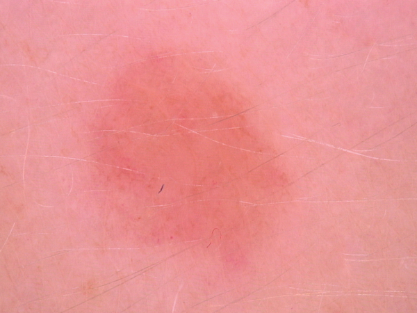
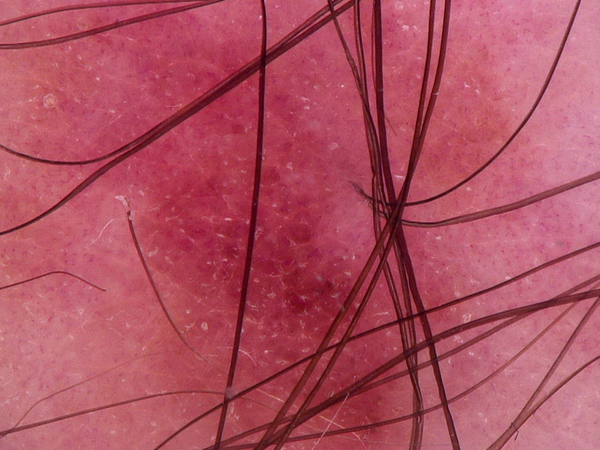
P23 classifies dermoscopy images across 7 skin disease classes using a zone-adaptive 3-engine ensemble and routes every case to one of four triage zones — from high-confidence benign to immediate specialist referral. A dedicated melanoma probability flag fires independently of the primary classification. A clinician always stays in command. Seven annotated demo cases span the full spectrum from high-confidence benign lesions to red-zone specialist deferral.
A confidence-zoned triage aid for dermoscopy review. Independent melanoma flag with specialist deferral on all red-zone and high-melanoma cases. Honest score, not a verdict.
🔬
Researchers
Validated ensemble metrics on HAM10000. Honest per-class accuracy reported as measured. MobileNetV2 transfer learning pipeline with zone-adaptive weighting. Full methodology, no cherry-picking.
🎓
Educators
A live classroom tool: seven annotated dermoscopy cases across 7 disease classes, confidence zones, melanoma flag discussion. Companion textbook and lab sheet for biomedical AI courses.
📖
Students
Explore how dermoscopy features translate to AI classification. See how an ensemble distributes probability across 7 classes and why a dermatologist must always review the result.
🏥
Hospital Planners
A demonstration of AI-augmented dermoscopy triage. PDPA-compliant. Zone protocol ensures borderline and melanoma-flag cases always reach a specialist, never a silent algorithmic verdict.
Three integrated capabilities
P23 combines confidence-zone triage with a dedicated melanoma safety flag and a seven-class dermoscopy classification engine. Each capability is designed to work together — classify the lesion, flag the melanoma risk, and know exactly when to defer.
Capability 1
🚦 Zone-Adaptive Confidence Triage
Every dermoscopy case is routed to one of four triage zones based on the ensemble’s confidence in its primary classification. Zone assignment drives the clinical recommendation — not just the label.
The zone-adaptive ensemble adjusts model weights according to the confidence zone, giving the most reliable engine a stronger voice when certainty is high.
4 zones · Zone-adaptive weights · Specialist deferral
Capability 2
⚠️ Independent Melanoma Flag
A dedicated melanoma probability flag fires independently of the primary classification result. When the ensemble assigns elevated melanoma probability — regardless of the top-ranked class — a specialist review warning is triggered.
This means a case classified as “Benign Keratosis” with elevated background melanoma probability still surfaces the warning. The primary label and the melanoma flag are reported separately; a clinician weighs both before any action.
The melanoma flag does not override the primary classification — it augments it.
Independent of top class · Always reported · Clinician decides
Capability 3
📊 7-Class Dermoscopy Classification
The 3-engine ensemble classifies across all seven skin disease classes from the HAM10000 / ISIC 2018 Task 3 benchmark. Per-class probabilities are returned for all seven classes — not just the top label.
The full probability distribution tells a clinician more than a single label: a 71% primary confidence with 18% on a malignant class is a different clinical situation than a 71% reading with the rest split evenly across benigns.
All seven demo cases are pre-loaded with real HAM10000 dermoscopy images and full probability bars.
7 classes · Full probability bars · HAM10000
Try it now — AEGIS SkinScan Demo Console
Seven dermoscopy cases from the HAM10000 dataset, each showing the full ensemble output: primary classification, confidence zone, melanoma flag status, and per-class probability bars across all seven disease classes. Select a case and explore why the system defers high-melanoma cases to a specialist rather than forcing a verdict.
All seven demo cases use HAM10000 / ISIC 2018 Task 3 dermoscopy images — open research dataset, consented and published. Never a clinical diagnosis. A qualified dermatologist must interpret all findings.
The four triage zones — confidence drives the clinical recommendation
Zone assignment is computed from ensemble confidence in the primary classification. The four zones map directly to clinical action: higher confidence enables a firmer recommendation, lower confidence triggers mandatory human review. The melanoma flag operates independently of zone assignment.
GREEN — High Confidence
Ensemble strongly supports the primary classification. Clinical recommendation can be acted on with the primary label as the leading signal. Melanoma flag still checked independently.
YELLOW — Mid Confidence
Ensemble reasonably supports the classification but with lower certainty. Dermatologist notification advised before any clinical decision. Melanoma flag reviewed carefully.
AMBER — Low Confidence
Ensemble uncertainty is elevated. Human review required before any clinical decision. Do not act on the primary label alone. Treat as equivalent to specialist consult.
RED — Defer. Specialist Required.
Ensemble confidence is insufficient for any clinical recommendation. Mandatory immediate specialist referral. No AI output from this system is sufficient to act on. Clinician must review.
The melanoma flag — the most important safety feature in P23. Melanoma is the deadliest form of skin cancer. The melanoma flag fires independently of the primary classification: a lesion classified as “Benign Keratosis” can still trigger the melanoma flag if the ensemble assigns elevated melanoma probability. When the flag fires, specialist review is always recommended, regardless of triage zone. This is a deliberate clinical design choice — a missed melanoma is a far greater harm than an unnecessary referral. A clinician always makes the final call.
The seven skin disease classes
AEGIS P23 classifies across the seven HAM10000 / ISIC 2018 Task 3 categories, spanning malignant, potentially malignant, and benign lesions. The full per-class probability distribution is returned for every case — not just the top label.
⚠️ Malignant
MEL — Melanoma
Deadliest skin cancer. Irregular pigmentation, atypical dermoscopic patterns. Any elevated melanoma probability triggers the independent melanoma flag regardless of zone.
⚠️ Malignant
BCC — Basal Cell Carcinoma
Most common skin cancer. Arborizing telangiectasia, leaf-like structures, rolled borders. High-confidence BCC classification triggers specialist referral recommendation.
🟡 Suspicious
AK — Actinic Keratoses
Pre-malignant lesion with potential for SCC progression. Scaly erythematous patch on sun-damaged skin. Dermatologist notification recommended in all zones.
🟡 Watch
VASC — Vascular Lesions
Haemangiomas, angiokeratomas, pyogenic granulomas. Red lacunae, irregular vessel patterns. Often classified with lower ensemble confidence — watch the zone carefully.
✅ Benign
NV — Melanocytic Nevi
Common mole. Symmetric pigment network, regular borders. Most frequent benign category in HAM10000. High-confidence NV classification is a reassuring signal.
✅ Benign
BKL — Benign Keratosis
Seborrhoeic keratosis, lichen planus-like keratosis. Milia-like cysts, comedo-like openings. Melanoma flag still checked even on high-confidence BKL results.
✅ Benign
DF — Dermatofibroma
Central white scar-like patch, peripheral pigmented network, positive pinch sign. Benign but often classified at lower confidence — mid-zone result is common for DF.
The biomedical engineering science
P23 is grounded in dermoscopy science, transfer learning theory, and clinical AI safety design. The feature extraction pipeline, ensemble architecture, and zone-adaptive weighting all derive from first principles — not arbitrary choices.
🩺 Dermoscopy and skin lesion classification
Dermoscopy (epiluminescence microscopy) illuminates sub-surface skin structures invisible to the naked eye — pigment network, vascular patterns, regression structures, and architectural disorder. These dermoscopic features encode clinically meaningful information about lesion biology. Melanoma, for example, shows irregular pigmentation, atypical vascular structures, and regression areas that are visible dermoscopically but not clinically. AEGIS P23 learns to read these features from the HAM10000 / ISIC 2018 Task 3 dataset — 13,611 dermoscopy images across 7 disease classes, the benchmark established by the International Skin Imaging Collaboration.
P23 uses MobileNetV2 pre-trained on ImageNet as the feature extractor — a lightweight convolutional architecture that captures rich visual representations from dermoscopy images without requiring millions of labelled training examples. The extracted feature representation is passed through a dimensionality reduction stage to produce a compact, discriminative feature vector. Three independently trained classifiers — Nu-SVC, SVM, and XGBoost — each learn from this feature space and vote on the classification. The zone-adaptive ensemble adjusts the voting weights based on confidence zone, ensuring the most reliable signal leads when certainty is high. The full per-class probability distribution is derived from ensemble output.
All engines trained and evaluated on distilled datasets drawn from HAM10000 / ISIC 2018 Task 3 under strict train/test separation. Validation methodology ensures no test image appears in training. Results reported exactly as measured — no inflation, no cherry-picking.
0.9131
AUC · High-confidence zone ensemble
91.24%
Accuracy · High-confidence zone cases
55.9%
Cases routed to high-confidence zone
Engine
Architecture
Validation Accuracy
Role
Nu-SVC
Nu-Support Vector Classifier
70.5%
Ensemble member — zone-adaptive vote
SVM
Support Vector Machine (RBF)
70.5%
Ensemble member — zone-adaptive vote
XGBoost
Gradient Boosted Trees
63.7%
Ensemble member — zone-adaptive vote
Honest note: individual model accuracy of 70.5% on a 7-class dermoscopy problem is a meaningful result on an imbalanced dataset with 13,611 images. The zone-adaptive ensemble substantially improves on this for the 55.9% of cases it classifies with high confidence (AUC 0.9131, accuracy 91.24%). The remaining 44.1% are routed to lower-confidence zones — not silently misclassified, but explicitly flagged for human review. That is the correct clinical design.
Zone-adaptive ensemble — the key architectural decision. Rather than using a fixed ensemble weighting for all cases, P23 adapts the engine weights according to the confidence zone. When the ensemble is most certain, the weights favour the engines that perform best in that regime. When uncertainty is higher, the weighting adjusts to be more conservative. This is why the high-confidence zone achieves AUC 0.9131 while the overall system correctly identifies and defers the harder cases rather than guessing.
Dataset integrity and data governance
P23 is trained and validated on a distilled research dataset derived from HAM10000 / ISIC 2018 Task 3 — a publicly available, consented dermoscopy image archive from the International Skin Imaging Collaboration. Raw patient dermoscopy images are not distributed in the public demo. The seven demo cases embedded in the public console are HAM10000 images released for open research use under their original dataset licence.
Illustration — AI-generated, decorative.
These apps demonstrate clinical AI systems capable of classification, early pre-emptive detection, and progressive augmented management. Because full AEGIS engines and datasets run to several gigabytes, only educational demo modes are hosted here — live classification on new patient dermoscopy images requires the AEGIS Desktop Application. Institutions wishing to contribute local data for engine refinement are welcome to reach out; we share progressive validation results with every contributing partner and operate in strict accordance with PDPA.
What institutional partners receive: technical report with full validation methodology · AEGIS Desktop Application licence · progressive model result updates as the engine board improves · co-attribution in peer-reviewed publications where applicable.
Ground rules for data contribution and institutional enquiry.
1. Not a medical device and not a diagnosis. AEGIS SkinScan is a research and educational demonstration. Nothing it outputs is medical advice, a diagnosis, or a substitute for a qualified dermatologist.
2. De-identified data only. Remove all PHI before contributing. Only submit data under your institution’s ethics approval and with informed consent.
3. Liability release. Contributing institutions agree to indemnify and hold harmless AEGIS, Dr Loh Kah Meng, and associated parties from any claims arising from use of or reliance on AEGIS outputs.
4. DUA required. A formal Data Use Agreement is required before any institutional data exchange proceeds.
5. PDPA compliance. All data handled under strict PDPA and applicable governance frameworks.